X-ray images with ORTHOPHOS XG units from Sirona are now easier to interpret than ever before. Software algorithms reduce metal artifacts in the DVT volume and improve image quality without increasing the radiation dose (MARS). In the area of 2D panoramic imaging, ASTRA provides brilliant, high-contrast images and the additional small exposure volumes and high-definition mode in ORTHOPHOS XG 3D allow even greater flexibility of use.
Sirona once again proves the lasting value of its ORTHOPHOS line with a comprehensive software package. The new features reduce noise and artifacts effectively. In addition, the package includes the option to switch to a smaller field of view with a resolution of 100 µm.
MARS reduces metal artifacts
The ORTHOPHOS XG 3D combination unit includes several improvements with "Release 2": Just like GALILEOS, ORTHOPHOS XG 3D now also works in 3D mode with MARS (Metal Artifact Reduction Software). MARS is an algorithm which automatically detects problematic metal artifacts and eliminates their shadowing and reflexes as much as possible. The result is a clear image which makes diagnosis easier for the dentist. MARS optimizes image quality without additional work or an increase in radiation dose. "Sirona is synonymous with responsibility in x-ray radiation. Therefore, it is our aim to obtain the best image quality with the lowest dose and a perfect workflow," says Wilhelm Schneider, Marketing Director of Imaging Systems at Sirona.
Optional high-definition mode for increased diagnostic reliability
There are cases, such as surgical interventions, in which a stronger radiation dose is required in order to ensure safety. An optional high-definition (HD) mode is now available for these kinds of treatments. ORTHOPHOS XG 3D produces 500 single exposures instead of 200 in a single cycle, from which the 3D x-ray image is computed. The resulting higher granularity and contrast makes it easier for the practitioner to make a diagnosis. The touch panel of the ORTHOPHOS XG 3D allows you to switch the HD mode on or off at any time.
Added volume for optimized endodontic treatments
In addition to the previous cylindrical image volume of 8 cm diameter x 8 cm height, ORTHOPHOS XG 3D can now depict a smaller volume of 5 cm diameter x 5.5 cm height. This smaller volume is also especially well suited for endodontics because the smaller section not only minimizes radiation exposure for the patient, but also speeds up the diagnostic process for the dentist. In addition, the new HD mode can be used with this smaller image volume if required. During this process, the voxel size automatically switches to 100 µm. This higher image resolution is then always indicated when the smallest details matter.
ASTRA suppresses image noise
All the new x-ray units in the ORTHOPHOS XG family work with a software algorithm, which greatly improves the image quality of 2D panoramic and ceph images. ASTRA (Anatomically STructured Reconstruction Algorithm) generates a clear image with higher contrasts, less noise and no problematic edge artifacts, thereby providing the ideal basis for a reliable diagnosis.
ORTHOPHOS XG 3D units can be upgraded with the "RELEASE 2" update package at no extra cost. ASTRA is not included in Release 2. Members of the software club Xwin Gold and Platinum receive ASTRA free of charge.
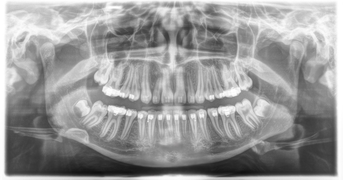
A great moment in x-ray technology : Brilliant image quality with MARS and ASTRA
InfoWeb Marketplace