Radiography 2015: New technology revolutionizes image quality with the smallest possible radiation dose
Sirona plans to present many innovations in the field of imaging systems at IDS 2015. New products and technologies not only deliver the highest possible image quality, but also support integrated digital solutions for efficient workflows in implantology, endodontics, orthodontics and prosthetics.
In order to make a reliable diagnosis in complicated cases, dentists need X-ray images of the highest quality, free of artifacts. This is ensured by innovative digital X-ray technology and the perfect interplay between hardware and software. Sirona has responded to the challenge with a completely updated product range for intraoral, 2D, and 3D radiography to be unveiled at this year's IDS trade fair. All Sirona X-ray equipment is fully supported by the new Sidexis 4 software for capturing, processing and archiving X-ray images.
Best 2D and 3D radiography with Orthophos SL
Besides the successful Orthophos XG, Sirona has also developed a new radiography system with innovative imaging technology that enables the sharpest possible imaging with the lowest possible X-ray dose: Orthophos SL is available as an upgradable 2D variant and as a 3D hybrid device with two volume sizes: 11x10 cm or 8x8 cm (particularly popular in general dental practices). The new direct conversion sensor converts the X-ray radiation directly into electrical pulses, not first into light. This results in reduced signal loss. The high yield of image data provides better image quality in panoramic images while also reducing the radiation dose. The reconstructive sharp layer process also generates several thousand individual projections in a single pass, shown from different angles, and reproducing the individual morphological conditions with pinpoint clarity. In 3D mode, many improvements such as metal artifact reduction and switchable HD mode enable outstanding image quality.
Radiography software that sets the standard
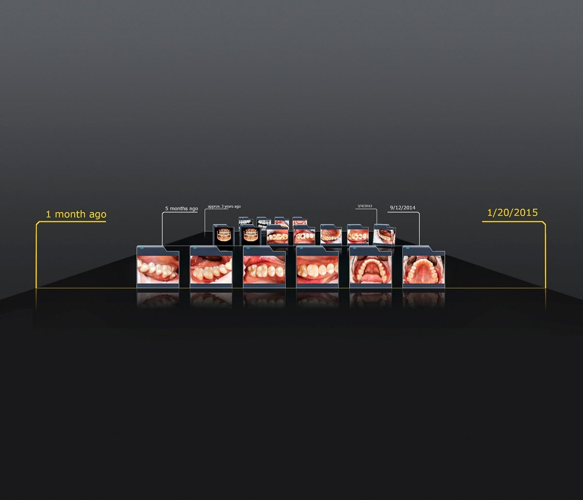
The images generated by this method can be optimally processed using the new Sidexis 4 radiography software. The program is characterized by its unrivaled workflow-oriented user interface and very easy handling. From the user interface, dentists can access all pertinent patient image data. Dentists thus have a full overview of the patient's treatment history. They can make cross references within the software, for example, compare intraoral and 3D images. A large number of interfaces enable Sidexis 4 to be connected to other systems, such as practice administration or the Teneo treatment center. The software suite can also be extended with plug-ins and applications to enable perfect coordination with integrated digital workflows, for simultaneous surgical and prosthetic planning of implants or to analyze the airways in patients with obstructive sleep apnea using Sicat Air.
Flexible entry to intraoral imaging
Sidexis 4 supports Sirona's full range of intraoral radiography systems: the Xios XG x-ray sensors, the Xios Scan X-ray scanner, and the latest generation of Sirona imaging plates. The Xios XG Supreme intraoral x-ray sensor offers a theoretical resolution of 33.3 line pairs per millimeter (Lp/mm), resulting in exceptional image quality. The sensor is very easy to use and patient friendly. It is available in three sensor sizes and has rounded edges, so it can be positioned well in the patient's mouth. Alternatively, in practices where the familiar workflow is to be retained, imaging plates can also be used. But instead of developing film, in this process they are digitized with a scanner. The user-friendly, low-maintenance XIOS Scan generates high resolution images with 22 Lp/mm. It can be easily connected to a PC and integrated in an existing practice network.
Digital radiography offers many advantages for intraoral imaging. With the aid of filters, image data can be optimized with sharpness, brightness and contrast to make complicated anatomical structures more visible. Handling digital x-ray images is also much simpler. The images are available immediately at the click of a mouse and do not have to be archived in a separate room where they have to be located for subsequent patient visits.