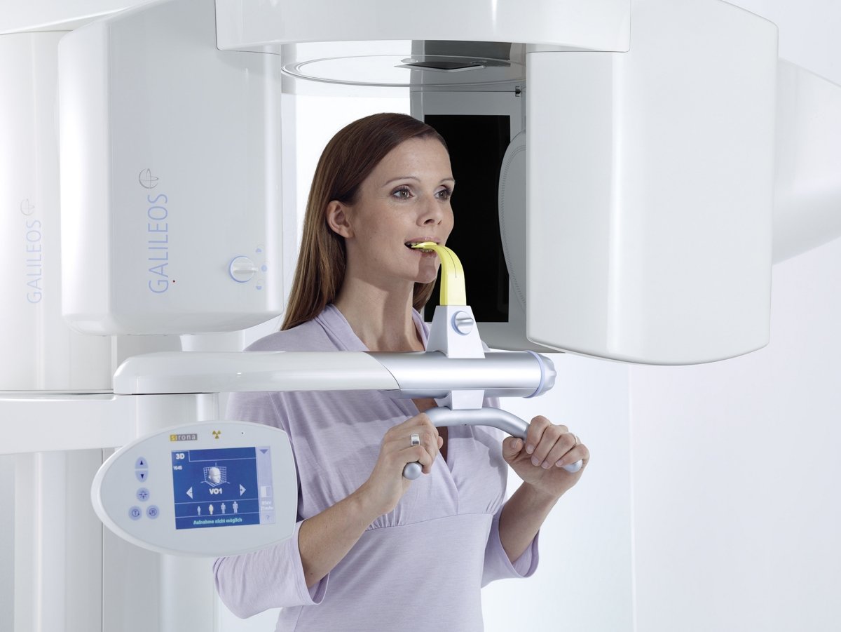
GALILEOS integrated face scanner brings the vision of the virtual patient one step closer. A new endodontic program for the ORTHOPHOS XG 3D.
Despite the rapid advances in digital dentistry, patients will still have to actually present themselves in person at the dental practice for treatment – no avatar can do that for them. And yet the prospect of the virtual patient is just around the corner. In assisting patient counselling and precise treatment planning, an avatar generated by omputer provides valuable assistance to the dentist and makes it easier for the patient to understand the proposed treatment. The basis for this is provided by the digital volume scans taken by the 3D-X-ray systems, GALILEOS und ORTHOPHOS XG 3D.
With its Integrated Face Scanning (IFS) Sirona has brought the prospect of the virtual patient a step closer. Thanks to the integration of a 3D scanner in GALILEOS, X-rays and surface anatomy scans can be taken simultaneously. The CBCT and IFS data are then superimposed with a very high degree of accuracy. The result is a lifelike depiction of the anatomical structures of the face, teeth and bones. The virtual mirror image formed in this way assists the dentist in planning treatment and makes it easier for the patient to understand the treatment proposed. Moreover, patients find it less disconcerting to look at their own face than an X-ray of their skull.
Precise virtual planning also forms the basis for safe and successful endodontic procedures. This is why Sirona has now added a special endodontic programme to its new hybrid X-ray unit ORTHOPHOS XG 3D. With both a collimated volume and high image resolution, the 3D programme has been designed to fully meet the requirements of root treatment. This hybrid unit has has been specially developed for general dental practices. It is based on the tried and tested ORTHOPHOS XG Plus and was launched on the market by Sirona at the end of 2010. It combines the advantages of two and three dimensional imaging extremely efficiently. Comprehensive panorama and cephalometric
X-ray programs minimise the level of radiation exposure. This new 3D function increases diagnostic security and, in combination with CEREC, opens up to the user new possibilities in the field of implantology.
"The more complex a dental procedure, the more important is clear communication with the patient," said Wilhelm Schneider, Marketing Director, Sirona Imaging Systems. "Three dimensional images make it easier for the patient to understand the proposed treatment and make it more likely they will accept it - the basis of successful treatment. That is why we continuously develop our X-ray systems further until the vision of the virtual patient becomes reality."
Virtual planning for optimal treatment – Sirona presents 3D X-ray innovations
InfoWeb Marketplace
